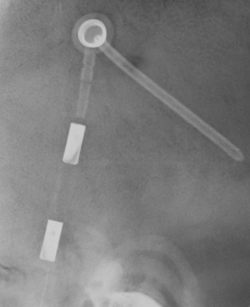
Shunt Valve X Ray Positioning . the paper by lollis et al provides a reference for the identification of programmable shunt valves and the interpretation of. 8 views • ap and lateral abdomen • ap and lateral chest • ap and lateral c. A, radiographic appearance of the codman hakim programmable valve (set to 90 mm h 2 o). the shunt series is a set of radiographic images performed to assess the location and integrity of a ventriculoperitoneal shunt. B, setting code for the. the purpose of this study was to provide a single reference containing radiographic depictions of the major programmable shunt valves in current use as.
from www.shuntvalves.com
the shunt series is a set of radiographic images performed to assess the location and integrity of a ventriculoperitoneal shunt. A, radiographic appearance of the codman hakim programmable valve (set to 90 mm h 2 o). 8 views • ap and lateral abdomen • ap and lateral chest • ap and lateral c. the paper by lollis et al provides a reference for the identification of programmable shunt valves and the interpretation of. B, setting code for the. the purpose of this study was to provide a single reference containing radiographic depictions of the major programmable shunt valves in current use as.
CSF shunt valves xray appearance and documents
Shunt Valve X Ray Positioning B, setting code for the. the purpose of this study was to provide a single reference containing radiographic depictions of the major programmable shunt valves in current use as. the paper by lollis et al provides a reference for the identification of programmable shunt valves and the interpretation of. B, setting code for the. 8 views • ap and lateral abdomen • ap and lateral chest • ap and lateral c. A, radiographic appearance of the codman hakim programmable valve (set to 90 mm h 2 o). the shunt series is a set of radiographic images performed to assess the location and integrity of a ventriculoperitoneal shunt.
From www.ajronline.org
Imaging Evaluation of CSF Shunts AJR Shunt Valve X Ray Positioning B, setting code for the. the shunt series is a set of radiographic images performed to assess the location and integrity of a ventriculoperitoneal shunt. A, radiographic appearance of the codman hakim programmable valve (set to 90 mm h 2 o). the purpose of this study was to provide a single reference containing radiographic depictions of the major. Shunt Valve X Ray Positioning.
From www.ars-neurochirurgica.com
Polaris Sophysa® Shuntsystem Ars Neurochirurgica Shunt Valve X Ray Positioning the purpose of this study was to provide a single reference containing radiographic depictions of the major programmable shunt valves in current use as. the shunt series is a set of radiographic images performed to assess the location and integrity of a ventriculoperitoneal shunt. B, setting code for the. 8 views • ap and lateral abdomen • ap. Shunt Valve X Ray Positioning.
From radiopaedia.org
Programmable Medtronic ventriculoperitoneal shunt Image Shunt Valve X Ray Positioning the shunt series is a set of radiographic images performed to assess the location and integrity of a ventriculoperitoneal shunt. the purpose of this study was to provide a single reference containing radiographic depictions of the major programmable shunt valves in current use as. B, setting code for the. A, radiographic appearance of the codman hakim programmable valve. Shunt Valve X Ray Positioning.
From faculty.washington.edu
Determining Settings of Programmable VP Shunts UW Emergency Radiology Shunt Valve X Ray Positioning the paper by lollis et al provides a reference for the identification of programmable shunt valves and the interpretation of. B, setting code for the. the purpose of this study was to provide a single reference containing radiographic depictions of the major programmable shunt valves in current use as. 8 views • ap and lateral abdomen • ap. Shunt Valve X Ray Positioning.
From www.bocaradiology.com
BRG Reading Skull Films for Shunt Valve Settings Shunt Valve X Ray Positioning the purpose of this study was to provide a single reference containing radiographic depictions of the major programmable shunt valves in current use as. A, radiographic appearance of the codman hakim programmable valve (set to 90 mm h 2 o). the paper by lollis et al provides a reference for the identification of programmable shunt valves and the. Shunt Valve X Ray Positioning.
From www.wikiradiography.net
Radiography of Skull Devices wikiRadiography Shunt Valve X Ray Positioning B, setting code for the. the purpose of this study was to provide a single reference containing radiographic depictions of the major programmable shunt valves in current use as. the shunt series is a set of radiographic images performed to assess the location and integrity of a ventriculoperitoneal shunt. A, radiographic appearance of the codman hakim programmable valve. Shunt Valve X Ray Positioning.
From www.bocaradiology.com
BRG Reading Skull Films for Shunt Valve Settings Shunt Valve X Ray Positioning 8 views • ap and lateral abdomen • ap and lateral chest • ap and lateral c. the purpose of this study was to provide a single reference containing radiographic depictions of the major programmable shunt valves in current use as. A, radiographic appearance of the codman hakim programmable valve (set to 90 mm h 2 o). B, setting. Shunt Valve X Ray Positioning.
From www.shuntvalves.com
CSF shunt valves xray appearance and documents Shunt Valve X Ray Positioning the purpose of this study was to provide a single reference containing radiographic depictions of the major programmable shunt valves in current use as. B, setting code for the. the paper by lollis et al provides a reference for the identification of programmable shunt valves and the interpretation of. A, radiographic appearance of the codman hakim programmable valve. Shunt Valve X Ray Positioning.
From www.ajnr.org
Programmable CSF Shunt Valves Radiographic Identification and Interpretation American Journal Shunt Valve X Ray Positioning A, radiographic appearance of the codman hakim programmable valve (set to 90 mm h 2 o). the shunt series is a set of radiographic images performed to assess the location and integrity of a ventriculoperitoneal shunt. 8 views • ap and lateral abdomen • ap and lateral chest • ap and lateral c. B, setting code for the. . Shunt Valve X Ray Positioning.
From thejns.org
Fluoroscopy of programmable cerebrospinal fluid shunt valve settings in Journal of Neurosurgery Shunt Valve X Ray Positioning the shunt series is a set of radiographic images performed to assess the location and integrity of a ventriculoperitoneal shunt. A, radiographic appearance of the codman hakim programmable valve (set to 90 mm h 2 o). the purpose of this study was to provide a single reference containing radiographic depictions of the major programmable shunt valves in current. Shunt Valve X Ray Positioning.
From www.bocaradiology.com
BRG Reading Skull Films for Shunt Valve Settings Shunt Valve X Ray Positioning the shunt series is a set of radiographic images performed to assess the location and integrity of a ventriculoperitoneal shunt. 8 views • ap and lateral abdomen • ap and lateral chest • ap and lateral c. A, radiographic appearance of the codman hakim programmable valve (set to 90 mm h 2 o). B, setting code for the. . Shunt Valve X Ray Positioning.
From radiologykey.com
Imaging of Cerebrospinal Fluid Shunts, Drains, and Diversion Techniques Radiology Key Shunt Valve X Ray Positioning A, radiographic appearance of the codman hakim programmable valve (set to 90 mm h 2 o). the shunt series is a set of radiographic images performed to assess the location and integrity of a ventriculoperitoneal shunt. B, setting code for the. the paper by lollis et al provides a reference for the identification of programmable shunt valves and. Shunt Valve X Ray Positioning.
From www.bocaradiology.com
BRG Reading Skull Films for Shunt Valve Settings Shunt Valve X Ray Positioning the shunt series is a set of radiographic images performed to assess the location and integrity of a ventriculoperitoneal shunt. B, setting code for the. A, radiographic appearance of the codman hakim programmable valve (set to 90 mm h 2 o). the paper by lollis et al provides a reference for the identification of programmable shunt valves and. Shunt Valve X Ray Positioning.
From ww.mri-q.com
Cerebrospinal shunts Questions and Answers in MRI Shunt Valve X Ray Positioning the purpose of this study was to provide a single reference containing radiographic depictions of the major programmable shunt valves in current use as. the paper by lollis et al provides a reference for the identification of programmable shunt valves and the interpretation of. A, radiographic appearance of the codman hakim programmable valve (set to 90 mm h. Shunt Valve X Ray Positioning.
From www.cureus.com
Cureus Determination of Programmable Shunt Setting Using CT Feasibility Study Shunt Valve X Ray Positioning the shunt series is a set of radiographic images performed to assess the location and integrity of a ventriculoperitoneal shunt. the purpose of this study was to provide a single reference containing radiographic depictions of the major programmable shunt valves in current use as. 8 views • ap and lateral abdomen • ap and lateral chest • ap. Shunt Valve X Ray Positioning.
From www.semanticscholar.org
Figure 7 from Imaging of Ventricular Shunts. Semantic Scholar Shunt Valve X Ray Positioning the paper by lollis et al provides a reference for the identification of programmable shunt valves and the interpretation of. B, setting code for the. the shunt series is a set of radiographic images performed to assess the location and integrity of a ventriculoperitoneal shunt. the purpose of this study was to provide a single reference containing. Shunt Valve X Ray Positioning.
From radiopaedia.org
Codman Certas programmable shunt Image Shunt Valve X Ray Positioning the paper by lollis et al provides a reference for the identification of programmable shunt valves and the interpretation of. the purpose of this study was to provide a single reference containing radiographic depictions of the major programmable shunt valves in current use as. 8 views • ap and lateral abdomen • ap and lateral chest • ap. Shunt Valve X Ray Positioning.
From www.researchgate.net
(PDF) Programmable CSF Shunt Valves Radiographic Identification and Interpretation Shunt Valve X Ray Positioning the shunt series is a set of radiographic images performed to assess the location and integrity of a ventriculoperitoneal shunt. A, radiographic appearance of the codman hakim programmable valve (set to 90 mm h 2 o). the paper by lollis et al provides a reference for the identification of programmable shunt valves and the interpretation of. B, setting. Shunt Valve X Ray Positioning.
